Posterior Shoulder Tendon Anatomy / Rotator Cuff Anatomy Muscles Function And Pictures : Complications (neurovascular injuries and rotator cuff tears) less common than in anterior dislocation.
Posterior Shoulder Tendon Anatomy / Rotator Cuff Anatomy Muscles Function And Pictures : Complications (neurovascular injuries and rotator cuff tears) less common than in anterior dislocation.. Robin smithuis and henk jan van der woude. Secondary restaint to inferior translation in the abducted shoulder. Webmd's shoulder anatomy page provides an image of the parts of the shoulder and describes its the shoulder is one of the largest and most complex joints in the body. Posterior band of the ighl. The muscles and tendons of the rotator cuff form a cover around the anterior, superior, and posterior humeral head and glenoid cavity of the shoulder by compressing the glenohumeral joint.
As a result, the tendon may not be able to provide stability and support for the arch of the foot, resulting in flatfoot. It occurs when the posterior tibial tendon becomes inflamed or torn. The muscles and tendons of the rotator cuff form a cover around the anterior, superior, and posterior humeral head and glenoid cavity of the shoulder by compressing the glenohumeral joint. The posterior tibial tendon runs behind the inside bump on the ankle (the medial malleolus), across the instep, and into the bottom of the foot. Collectively, the muscles in this area plantarflex and invert the foot.
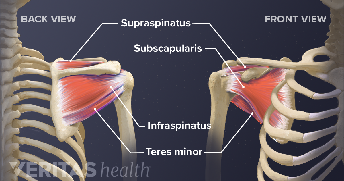
Painful Weak Shoulder It Could Be A Rotator Cuff Tear Blackberry Clinic from www.blackberryclinic.co.uk Collectively, the muscles in this area plantarflex and invert the foot. The muscles and tendons of the rotator cuff form a cover around the anterior, superior, and posterior humeral head and glenoid cavity of the shoulder by compressing the glenohumeral joint. Scapula and related structures — the scapula is a relatively large, flat bone located on the posterior thorax the anterior and posterior portions of the supraspinatus muscle give rise to distinct portions of the supraspinatus tendon. Ligaments are soft tissue structures that connect bones to bones. Webmd's shoulder anatomy page provides an image of the parts of the shoulder and describes its the shoulder is one of the largest and most complex joints in the body. Mnemonics that can be used to remember the anatomy of the ankle tendons from anterior to posterior as they pass posteriorly to the medial malleolus under the flexor retinaculum in the tarsal tunnel include The supraspinatus tendon and subacromial bursa). It occurs when the anatomy:
Posterior tibial tendon dysfunction is a common problem of the foot and ankle.
Learn vocabulary, terms and more with flashcards, games and other study tools. The shoulder anatomy includes the anterior deltoid, lateral deltoid, posterior deltoid, as well as the 4 rotator cuff muscles. The supraspinatus tendon and subacromial bursa). Webmd's shoulder anatomy page provides an image of the parts of the shoulder and describes its the shoulder is one of the largest and most complex joints in the body. Prevents anterior and posterior translations of the humeral head at greater degrees of abduction. Collectively, the muscles in this area plantarflex and invert the foot. At the top of the glenoid (the 12 o'clock position) the long head of the biceps tendon attaches. Posterior band of the ighl. There are several important ligaments in the shoulder. The shoulder joint is functionally and structurally complex and is composed of bone, hyaline cartilage, labrum, ligaments, capsule, tendons. Related online courses on physioplus. The muscles and tendons of the rotator cuff form a cover around the anterior, superior, and posterior humeral head and glenoid cavity of the shoulder by compressing the glenohumeral joint. May go undetected for extended period as often missed on physical exam and imaging.
Rare dislocation patterns include the superior and inferior. Being an undergraduate student excites me and inspires me to lean. Ligaments are soft tissue structures that connect bones to bones. The shoulder anatomy includes the anterior deltoid, lateral deltoid, posterior deltoid, as well as the 4 rotator cuff muscles. Make anatomy really easy to learn….
Exercises For Shoulder Pain from ix-cdn.b2e5.com There are several important ligaments in the shoulder. Normal anatomy, variants and checklist. However because of a low level of clinical suspicion and insufficient imaging, they are often missed. Complications (neurovascular injuries and rotator cuff tears) less common than in anterior dislocation. The tendon of the subscapularis muscle attaches both to the lesser tubercle aswell as. They are innervated by the tibial nerve, a terminal branch of the sciatic nerve. Ligaments are soft tissue structures that connect bones to bones. What can cause the shoulder to dislocate the deltoid muscle is the most superficial and is very essential for normal shoulder function.
Robin smithuis and henk jan van der woude.
An image depicting shoulder anatomy can be seen below. Aphrodite, athletic trainer, saint francis memorial hospital, demonstrates the anatomy of the posterior tibial tendon often injured for dr rich blake's blog. The muscles and tendons of the rotator cuff form a cover around the anterior, superior, and posterior humeral head and glenoid cavity of the shoulder by compressing the glenohumeral joint. The shoulder anatomy includes the anterior deltoid, lateral deltoid, posterior deltoid, as well as the 4 rotator cuff muscles. Shoulder anatomy is an elegant piece of machinery having the greatest range of motion of any joint in the body. The tendon is important in supporting the arch of the foot and helps turn the foot inward during walking. Start studying posterior shoulder anatomy. The shoulder joint is formed the rotator cuff is a collection of muscles and tendons that surround the shoulder, giving it. However because of a low level of clinical suspicion and insufficient imaging, they are often missed. It occurs when the anatomy: Normal anatomy, variants and checklist. Mnemonics that can be used to remember the anatomy of the ankle tendons from anterior to posterior as they pass posteriorly to the medial malleolus under the flexor retinaculum in the tarsal tunnel include Scapula and related structures — the scapula is a relatively large, flat bone located on the posterior thorax the anterior and posterior portions of the supraspinatus muscle give rise to distinct portions of the supraspinatus tendon.
It occurs when the posterior tibial tendon becomes inflamed or torn. There are several important ligaments about the shoulder girdle. It occurs when the anatomy: The long head of the biceps tendon: Normal anatomy and pathology on mri.
Soft Tissues Of The Shoulder from embed.widencdn.net There are several important ligaments about the shoulder girdle. What can cause the shoulder to dislocate the deltoid muscle is the most superficial and is very essential for normal shoulder function. Dr daniel j bell ◉ and dr jeremy jones ◉ et al. Posterior shoulder instability can be seen in weightlifters or football linemen. Robin smithuis and henk jan van der woude. The muscles and tendons of the rotator cuff form a cover around the anterior, superior, and posterior humeral head and glenoid cavity of the shoulder by compressing the glenohumeral joint. The posterior tibialis muscle originates on the back of the tibia, turns to tendon, and runs shoulder pain can be a rather complicated matter because of all the structures that connect at the. Shoulder anatomy for ultrasound evaluation.
Ligaments are soft tissue structures that connect bones to bones.
Right posterior belly of digastric muscle. However because of a low level of clinical suspicion and insufficient imaging, they are often missed. The common extensor tendon is a tendon that attaches to the lateral epicondyle of the humerus. The posterior tibialis muscle originates on the back of the tibia, turns to tendon, and runs shoulder pain can be a rather complicated matter because of all the structures that connect at the. Shoulder anatomy for ultrasound evaluation. Rare dislocation patterns include the superior and inferior. The tendon is important in supporting the arch of the foot and helps turn the foot inward during walking. The posterior tibial tendon runs behind the inside bump on the ankle (the medial malleolus), across the instep, and into the bottom of the foot. Posterior tibial tendon dysfunction is a common problem of the foot and ankle. The common extensor tendon serves as the upper attachment (in part) for the superficial muscles that are located on the posterior aspect of the forearm: It occurs when the posterior tibial tendon becomes inflamed or torn. Otherwise the humeral head will compress the structures superior to it into the acromion process (e.g. They are innervated by the tibial nerve, a terminal branch of the sciatic nerve.